For decades the focus of neuroscience was on neurons, but as research techniques and technology progresses we are beginning to understand the brain as never before. The importance of neurons cannot be downplayed, and yet scientists are coming to realise that there are many other elements involved in brain development.
Cilia are slender, hair-like organelles found on the surface of most cells. There are several conditions known to medical science which occur as a result of ciliary defects (e.g. scoliosis) but researchers at the Norwegian University of Science and Technology have now begun to investigate the role of cilia in healthy brain development.
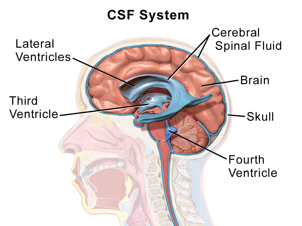
When cilia are functioning correctly, they beat in a coordinated rhythm to generate a directional fluid flow. Within the central nervous system (CNS), this mechanism generates the flow of cerebral spinal fluid (CSF) which transports nutrients and signaling molecules around the CNS and removes waste products (Del Bigio, 2010). Research also indicates that CSF flow may support neural stem cell proliferation and directional neuronal migration (Petrik et al, 2018; Sawamoto et al, 2006).
Previous research has indicated that fluid flow generated by cilia in the brain ventricles (interconnected fluid-filled chambers in the brain) may be regulated by the circadian rhythm and brain-derived neuropeptides. It has also been demonstrated that CSF composition varies across brain ventricles (Faubel et al, 2016; Conductier et al, 2013, Lun et al, 2015; Lun et al, 2015). All together this suggests that the nervous system can regulate the compartmentalization of CSF within the brain, but the specifics of how cilia might direct this CSF flow remain unclear.
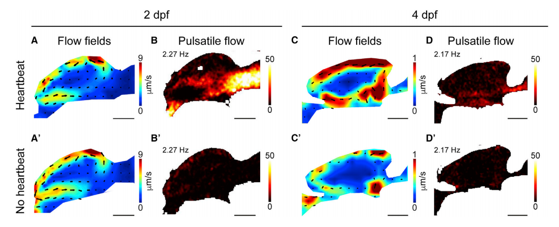
In order to investigate this mechanism further, Nathalie Jurisch-Yaksi and her team studied the trasparent brains of zebrafish larvae. Using cilia markers such as Foxj1, they measured ciliary beating in 2- to 4-day old larvae by light-sheet microscopy. Then then imaged the larvae brains to determine how ciliary beating might impact CSF flow.
The team found that CSF flow is generated by multiple parameters; ciliary beating strongly impacts near-wall flow, but heartbeat and body contractions also impact how CSF moves around the ventricles. Cilia compartmentalize the CSF flow by establishing ventricular boundaries, meaning very little CSF is exchanged at the boundary borders. Despite this, cilial beating was shown to be vital for keeping ventricular ducts between each border open; the researchers showed that interrupting cilia can disrupt ventricular development and reduce fluid coupling between brain ventricles.
This research provides an early indication of how cilia may play a role in brain functioning, however the comparison between zebrafish and human cilia cannot yet be established. The next step for the research team is to investigate what happens when cilia functioning is interrupted or manipulated, to better understand their influence on neural systems. For instance, the team question if circadiam rhythms might be altered if CSF flow is changed.

The research paper associated with this article has been published in Current Biology and Open Access. The paper can be downloaded here.
References
- Del Bigio, M.R. (2010). Ependymal cells: biology and pathology. Acta Neuropathol. 119, 55–73.
- Petrik, D., Myoga, M.H., Grade, S., Gerkau, N.J., Pusch, M., Rose, C.R., Grothe, B., and Go¨ tz, M. (2018). Epithelial sodium channel regulates adult neural stem cell proliferation in a flow-dependent manner. Cell Stem Cell 22, 865–878.e8.
- Sawamoto, K., Wichterle, H., Gonzalez-Perez, O., Cholfin, J.A., Yamada, M., Spassky, N., Murcia, N.S., Garcia-Verdugo, J.M., Marin, O., Rubenstein, J.L., et al. (2006). New neurons follow the flow of cerebrospinal fluid in the adult brain. Science 311, 629–632.
- Faubel, R., Westendorf, C., Bodenschatz, E., and Eichele, G. (2016). Ciliabased flow network in the brain ventricles. Science 353, 176–178.
- Conductier, G., Brau, F., Viola, A., Langlet, F., Ramkumar, N., Dehouck, B., Lemaire, T., Chapot, R., Lucas, L., Rove`re, C., et al. (2013). Melaninconcentrating hormone regulates beat frequency of ependymal cilia and ventricular volume. Nat. Neurosci. 16, 845–847.
- Lun,M.P., Johnson, M.B., Broadbelt, K.G.,Watanabe,M., Kang, Y.J., Chau, K.F., Springel, M.W., Malesz, A., Sousa, A.M., Pletikos, M., et al. (2015). Spatially heterogeneous choroid plexus transcriptomes encode positional identity and contribute to regional CSF production. J. Neurosci. 35, 4903– 4916.
- Lun, M.P., Monuki, E.S., and Lehtinen, M.K. (2015). Development and functions of the choroid plexus-cerebrospinal fluid system.Nat. Rev. Neurosci. 2015; 16: 445-457

